A typical radiology worklist no longer looks the way it did a few years ago. As studies populate the queue, AI-enabled tools may automatically flag suspected intracranial hemorrhage, highlight pulmonary nodules or prioritize cases based on predicted acuity. Measurements that once required manual input—lesion size, volumetrics or ejection fraction estimates—are now pre-populated, allowing you to verify rather than generate them from scratch.
Radiology is the most AI-active medical field, with more than 76% of FDA-cleared algorithms falling under medical imaging, according to the Radiological Society of North America (RSNA). AI in radiology automates measurements and integrates decision support directly into picture archiving and communication systems (PACS).
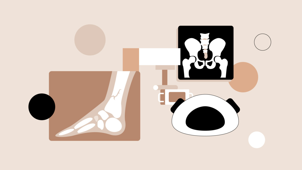
This article provides a peer-driven analysis of artificial intelligence in radiology today. Explore how clinicians use it, their top concerns and how technology will likely evolve from here.
What is artificial intelligence in radiology?
Artificial intelligence in radiology applies deep learning or other forms of machine learning (ML) to review medical images to support clinical decision-making.
If you need a refresher on AI terms, ML is a form of AI referring to systems that can learn from and be enhanced by data automatically. Deep learning is a subset of ML that shines at analyzing data on a large scale. Deep learning programs can rely on different types of algorithms, such as Convolution Neural Network (CNN). CNN is typically used in image and video recognition, and it’s commonly used in radiology AI.
Current top applications of AI in radiology have meaningful clinical traction in high-volume areas. Modalities like CT, MRI, X-ray, mammography and ultrasound feature the most mature AI applications due to their high imaging volume and standardized data availability. “AI’s strength in radiology currently is in imaging acquisition,” writes one radiology resident in the Sermo community. “It has decreased scan times and improved image quality, allowing for less patient exposure to radiation and increased availability of MRI scanners.”
Key applications of AI in radiology
AI diagnostic imaging tools are already embedded in daily diagnostic work. Here are four AI medical imaging examples you may encounter:
Triage and prioritization
AI flags critical findings like intracranial hemorrhage, pulmonary embolism or pneumothorax and moves them to the top of the worklist. This queue management is a high-impact application. Tools like Viz.ai or Aidoc for critical findings can reduce how long it takes you to interpret time-sensitive pathologies.
Detection (CAD)
Deep learning-based computer-aided detection (CAD) improves upon older systems that often produced distracting false positives. Current applications in mammography, lung nodule detection and fracture identification can highlight subtle morphological changes. While false positives still occur, modern algorithms are more refined.
Quantification
AI excels at tedious measurements like volumetric tumor tracking, cardiac function metrics and brain atrophy quantification. Framing this as “AI vs radiologist” would be misleading. This technology does not replace interpretation; it automates measurement grunt work so doctors can focus on complex synthesis.
Workflow automation
Radiology workflow automation saves time on non-interpretive tasks. Algorithms manage protocol selection, hanging protocols, auto-population of structured reports and intelligent routing. These practical tools enable faster report turnaround and better case flow.
Benefits of AI in radiology
The benefits of AI in radiology include more efficient workflows and reduced errors in high-volume tasks. You can think of AI as a safety net that supports your skills as a radiologist.
Accelerating image interpretation and reporting
AI speeds interpretation by pre-populating measurements, highlighting regions of interest and triaging critical cases. A chest X-ray AI can flag suspected pneumothorax so that you can act rapidly.
Improving detection of subtle or early-stage abnormalities
Deep learning models detect subtle texture changes that are difficult for humans to perceive consistently. This could mean identifying small pulmonary nodules on CT, microcalcifications on mammography or subtle fractures on plain film to improve sensitivity for early disease.
Reducing diagnostic errors or misses
By serving as a systematic second reader, AI reduces oversight errors. False negatives may fall when algorithms screen every image and call attention to borderline findings. That said, AI can produce false positives or false negatives too. “AI is a useful backup in radiology but should never be a primary reader,” cautions a radiologist on Sermo.
Enhancing workflow efficiency and case prioritization
Workflow AI automates repetitive tasks and integrates with voice recognition. This enables faster report turnaround and better case flow for the entire department.
Quantification and predictive analytics
Automated segmentation makes tumor volumes, ejection fraction and nodule sizing reproducible and auditable. This reduces interobserver variability and frees radiologists to concentrate on unexpected findings.
Expanding access in low-resource or remote settings
In areas with radiologist shortages, validated AI tools provide basic triage. This enables remote clinicians to identify urgent pathology and prioritize teleradiology workflows.
Extending decision support to pathology
AI augments pathology workflows by quantifying biomarkers, grading tumors and isolating candidate regions for immunohistochemistry. These decision-support outputs produce context-aware recommendations when integrated with clinical data.

How accurate is AI in radiology?
Research suggests that AI tools can help improve diagnostic accuracy, though they aren’t perfect. One study comparing an AI-based reporting platform with the traditional dictation method found that reporting time was faster and accuracy was higher when radiologists used AI-generated reports. However, the authors noted that the AI-generated reports did have occasional errors that a radiologist would need to pick up on when reading the reports.
AI radiology accuracy depends on data quality and the clinical environment. AI models are only as good as their training data. A tool trained at an academic medical center may not perform the same way at a community hospital using different equipment. This dataset shift means local validation is always necessary. Every tool is a decision support system—requiring physician oversight.
Some radiology AI tools are tuned for high sensitivity to avoid missing findings, which could produce more false positives. For example, a tool that flags 15 possible pulmonary embolisms per shift when only two are real creates alert fatigue, which can paradoxically reduce attention to true positives.
Physicians’ concerns about AI integration in radiology
Despite the promise, the risks of AI in radiology cause valid physician anxieties. “I am not a big fan of AI,” one emergency medicine doctor writes. “Something is inherently wrong when humans seek comfort from a machine.” These are some of the most common concerns surrounding the use of AI in radiology:
Liability and accountability in case of errors
When an AI-assisted report misses a diagnosis, it raises the question of whether the physician or the technology is responsible. Physicians worry that unclear liability will shift legal heat onto clinicians. “Also, it is hard to sue a machine, but far easier to sue a human being, hence patients will always prefer humans to machines,” asserts a pathologist on Sermo.
Accuracy and reliability of AI-generated outputs
As mentioned, AI can produce false positives or false negatives, so issues can arise if physicians place too much trust in the tools rather than remaining critical. “It’s incomprehensible to be carried away by something that isn’t proven,” writes one general practitioner on Sermo. One review of 83 existing studies found no significant performance difference between AI models and physicians, but the authors noted that accuracy varies by model.
Transparency of algorithms (the black box problem)
“Black box” AI refers to models that make predictions without transparent reasoning. Physicians need explainable outputs to assess when to trust an algorithm, so explainable AI (which reveals how it arrived at a conclusion) is preferable.
Reduced physician input or loss of clinical autonomy
Overreliance on AI could deskill trainees or normalize deferring to algorithmic outputs. “AI is useful for consulting but it cannot turn into the main decision-making tool,” a dermatologist on Sermo warns their colleagues.
Job displacement in the long term
The question of whether AI will replace radiologists altogether has proven controversial. The consensus among radiology leadership has been no, however AI’s role is evolving. Mitchell H. Katz, MD, CEO of the largest public health system in the U.S., said “we could replace a great deal of radiologists with AI at this moment,” after suggesting relying on AI for first reads of mammograms during a recent panel. On the other hand, recent investigations from CNN and The New York Times concluded that AI is positioned to enhance, but not replace, jobs in radiology.
Training needs
Practical AI literacy includes understanding key model performance metrics like sensitivity and specificity. Issues could arise if physicians aren’t familiar with sources of bias and model validation techniques.
Additional practical concerns
Physicians highlight the cybersecurity risks of connected AI services and the regulatory complexity of using continuously learning systems. Deep learning models reuses patient data, which raises the question of whether patients need to provide informed consent more than once, according to the authors of one study. AI devices can be vulnerable to threats like ransomware, they noted.
Challenges of implementing AI in radiology
To deploy radiology AI tools, you may have to overcome a few practical barriers.
Cost
AI tools carry licensing fees, infrastructure requirements and ongoing maintenance costs. For many community radiology practices operating on tight margins, the return on investment is not straightforward.
Integration (PACS/EHR)
AI tools need to integrate seamlessly into existing PACS workflows. They must receive images, process them and return results without disrupting established routines. Poor DICOM compatibility is a common reason promising tools fail adoption.
Training requirements
Radiologists and technologists need training to interpret AI outputs critically. You’ll benefit from understanding a tool’s known limitations and failure modes in addition to understanding where it excels. Traditional radiology, which focuses “heavily on mastering the nuances of image interpretation” will no longer be sufficient as AI tools become more widespread, the authors of one study argued. Radiologists will additionally need to learn “a broad spectrum of medical data, including complex non-imaging information such as genomic sequences, proteomic analyses, and advanced immunological markers,” in diagnostic decisions, they wrote.
Regulatory complexity
The regulatory landscape for medical devices is evolving. In the U.S., FDA 510(k) clearance and post-market surveillance requirements mean that algorithm updates can trigger new regulatory reviews. This is particularly relevant for AI tools that are continuously learning or being iteratively improved, as even small changes to an algorithm’s performance or intended use may require additional validation and regulatory approval. Health systems and radiology leaders are responsible for the ongoing compliance, monitoring and documentation required to safely maintain these tools.
The future role of radiologists and pathologists in an AI world
As AI capabilities mature, the role of radiologists and pathologists is likely to shift. Rather than focusing primarily on first-pass detection, diagnostic specialists could increasingly operate as integrators of information, synthesizing imaging or histopathologic findings with clinical context, prior studies and evolving patient data to guide management decisions.
AI systems are well-suited to repetitive, high-volume tasks such as identifying common abnormalities or generating standardized measurements. In contrast, radiologists and pathologists will continue to lead in areas where nuance matters: interpreting atypical presentations, reconciling discordant findings, and making judgment calls in complex or uncertain cases. These higher-order interpretive tasks remain difficult to automate reliably.
At the same time, physicians are taking on a growing role in the validation and oversight of AI tools. This includes participating in model selection, evaluating performance across diverse patient populations, monitoring for drift over time, and ensuring that outputs remain clinically meaningful. In many practices, this work is becoming part of routine quality assurance, requiring ongoing physician engagement rather than one-time implementation.
Physician responsibilities in an AI-enabled radiology practice
As AI becomes embedded in radiology workflows, its safe and effective use depends heavily on physician oversight. Radiologists can benefit from fluency in clinical AI literacy—not only understanding how to use these tools, but also how they generate outputs, where they may fail, and how they should (and should not) influence clinical decision-making. Radiologists remain ultimately accountable for the accuracy of the interpretation and the final report, regardless of whether they use AI-assisted tools.
These responsibilities align with broader ethical guidance from U.S. medical organizations. For instance, the American Medical Association (AMA) emphasizes that AI should augment, not replace, clinical judgment, and that physicians must understand the tools they use and remain accountable for patient care decisions. Similarly, the RSNA has highlighted the importance of transparency, validation, and ongoing performance monitoring in clinical AI deployment.
Shaping the future of diagnostic medicine with AI
Artificial intelligence is constantly evolving. Here’s how you can stay informed about new developments:
Weigh in on AI’s impact on diagnostics
Sermo is playing a critical role in this transition. As a private community for physicians, it can help you vet vendor claims, surface real-world learnings and shape ethical debates. Join the conversation to exchange real-world evaluations and collaborate on best practices.
Get The Sermo Roundup
Stay ahead with The Sermo Roundup newsletter. Receive your regular dose of peer insights, practical guides and trends shaping the medical profession.
How does AI affect your specialty?
You can strengthen your standards by engaging in peer discussions and sharing validation data. Join Sermo to see how your peers are navigating these technological shifts.